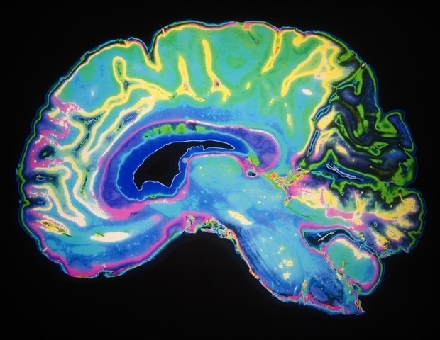
In experiments with healthy volunteers undergoing functional MRI imaging, scientists have increased activity in two regions of the brain that work together to react, and possibly regulate, when the brain feels “tired” and leaves either and continues mental efforts.
The experiments designed to help detect various aspects of brain fatigue, can provide physicians a way to better evaluate and treat them who experience mental exhaustion, including depression and post-tract stress disorder (PTSD), scientists say.
A report on NIH-funded study was published online on 11 June Neuroscience journal18 female and 10 male healthy adult volunteers extended the results to use their memory.
Our lab focuses on how [our minds] Produce value for effort. We understand less about biology of cognitive functions, including memory and recall, we do about physical functions, even though both include a lot of efforts. ,
Vikram Chib, PhD, Associate Professor of Biomedical Engineering at Johns Hopkins University School of Medicine and Research Scientist at Kennedy Criser Institute
Really, Chib says, scientists know that cognitive functions are tired, and relatively low why and how such fatigue develops and plays in the brain.
At the age of 21 to 29, 28 study participants were paid $ 50 to participate in the study, and they were told that they could receive additional payments based on their performance and options. All participants received a baseline MRI scan before the experiment begins.
His working memory tests, which were involved in a screen, on a screen, on a screen, on a screen, remembering a series of letters, remembering the position of some letters. A letter was in a series of letters, it was as hard as it was to remember its position, increasing cognitive efforts. Participants were reacted to their performance after opportunities to receive growing payments ($ 1-$ 8) with each test and more difficult recall practice. Participants were also asked to self-by-rate the level of cognitive fatigue before and after each test.
Overall, the results of the test increased activity and connectivity in two brain regions when participants reported cognitive fatigue: the right insula, the deeper into the brain is associated with an area that is associated with the feelings of fatigue, and the dorsal lateral prefrontal cortex, the fields on both sides of the brain control the memory. For each participant, during cognitive fatigue, the activity in both brain locations increased more than double the level of baseline measurement taken before starting tests.
“Our study was designed to inspire cognitive fatigue and to see how people feel tired, when they feel tired, as well as identifying places in the brain where these decisions are made,” Chib says.
In particular, the members of the Chib and his research team Grace Steward and Vivian Looi found that the participants should have financial incentive to increase cognitive efforts at a high level, suggests that external incentives induce such efforts.
“This result was not completely surprising, given our previous work in physical effort,” Chib is called.
Says Chib, “The two regions of the brain are working together to avoid more cognitive efforts until more encouragement is offered. However, cognitive fatigue may have an discrepancy between perceptions and the human brain is really able to do,” Chib says.
Fatigue is associated with many neurological conditions, including PTSD and depression, Chib says. “Now that we have identified some nerve circuits for cognitive efforts in healthy people, we need to see how fatigue appears in the minds of these conditions,” they say.
Chib says that it may be possible to use drug or cognitive behavior therapy to combat cognitive fatigue, and the current study may be a framework to clearly classify the current study cognitive fatigue using decisions and functional MRIs.
The functional MRI uses blood flow to measure the broader areas of activity in the brain; However, it directly measures neuron activation, nor more subtle nuances in brain activity.
“This study was done in an MRI scanner and with very specific cognitive functions. It would be important to see how these results normalize for other cognitive efforts and real -world functions,” Chib says.
The funding for research was provided by the National Institute of Health (R01HD097619, R01MH119086).
Source:
Journal reference:
Steward, G. At al. (2025). Its effect on neurobiology and effort-based options of cognitive fatigue. Journal of neurosciences, doi.org/10.1523/jneurosci.1612-24.2025,


